

Microscopes are an extremely useful tool to study really small objects. The instrument is almost synonymous with many scientific fields and is considered one of the greatest inventions by humanity. Constant research efforts to improve microscopes are currently being undertaken worldwide to meet the various demands that have arisen as different fields have advanced through the decades. One of the big problems in medicine has been the problem of non-invasive observation. We need to see what is going inside a patient for many different reasons; being able to do this without actually having to cut them up is a big plus.
The motivation for developing such techniques comes from the fact that some of the more complicated organs like brains are very difficult to operate on. Other problems include the scattering of light due to bone structures and the risk of tissue damage. An invention has come up that might be able to address all of these issues. Researchers have found a way to create a clear image from the scattered light emitted from a laser after it has passed through a thick layer of bone, like the skull. The microscope will make it possible to investigate living tissues that lie beyond thick bone layers with ease.
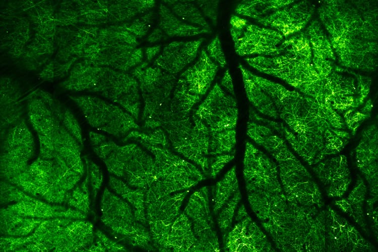
Other techniques before this one have captured images of brain tissues of mice, but most of them require cutting and opening up the skull. One method that doesn’t do so is called three-photon microscopy, and it uses a longer wavelength and a special gel to see through the bone. However, this method can only see so deep into the tissue. Another drawback is the combination of frequencies that can potentially damage fragile biological structures. The new microscope used computational adaptive optics to create the first-ever high-resolution image of mouse neurons from outside the skull.
The new invention is called laser-scanning reflection-matrix microscopy (LS-RMM); it works by not only detecting light scattering at the depth being imaged but also by getting a complete input-output response to the light-medium interaction, also known as the reflection-matrix. The power of this technique lies in the fact that it utilizes the maximum number of photons that have been scattered due to the different obstacles in the path of the light beam. All this led to the generation of the picture of the neurons inside the mouse’s brain. The new technique, however, requires a great deal of computing power to process all the data that it collects.
The potential use of this technique is very big in its scope. The new technique could help reveal more about the roles and functions of biological structures as it can help us see it in its natural living context. It might also prove helpful in early disease diagnosis and expedite neuroscience research. This might be the beginning of something very exciting.
Further Reading: