

The annual photography competition brings to light the smallest marvels that exist on Earth. It makes us realize how exquisite our planet and its creatures are. A beautiful world exists on our own Earth which is invisible to our naked eyes.
Development biologists Daniel Castranova, Bakary Samasa, and Brant Weinstein found this exquisiteness inside a zebrafish. They captured a stunning photograph of a young zebrafish while working in Weinstein’s lab at the National Institutes of Health in Bethesda, Md., illuminating the never before seen parts of the lymphatic system.
The picture was taken during research to find out whether zebrafish have lymphatic vessels inside their skulls. The lymphatic system is useful in clearing toxins and wastes from the body. Previously, researchers believed that only mammals have this structure close to their brain.
In the preliminary research published in May in bioRxiv.org, the team found that zebrafish have these vessels too. They used fish which were genetically modified. In those genetically modified fish, lymphatic vessels gave a fluorescent orange colour, and their skeletons and scales gave a blue glow under certain conditions.
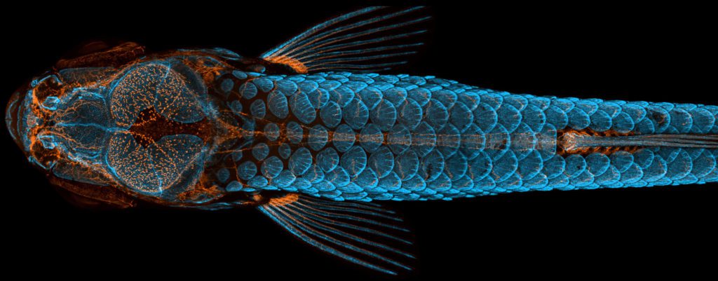
According to Castranova, since fish are easier to raise and image in labs than mammals, their research could help scientists study the role of the brain’s lymphatic system more easily in neurological diseases like brain cancer or Alzheimer’s.
They took a composite of 350 images with a confocal microscope on a busy working day. Castranova says, “I never even looked at the picture for a couple of weeks, and then when I looked at it at some point post-data processing, I was like ‘Wow.’”
The judges for the 2020 Nikon Small World photography competition announced this photo to be the winner. It received first place in the 46th annual photography competition, the results of which were announced on October 13th.
The second and third winners of the photography competition
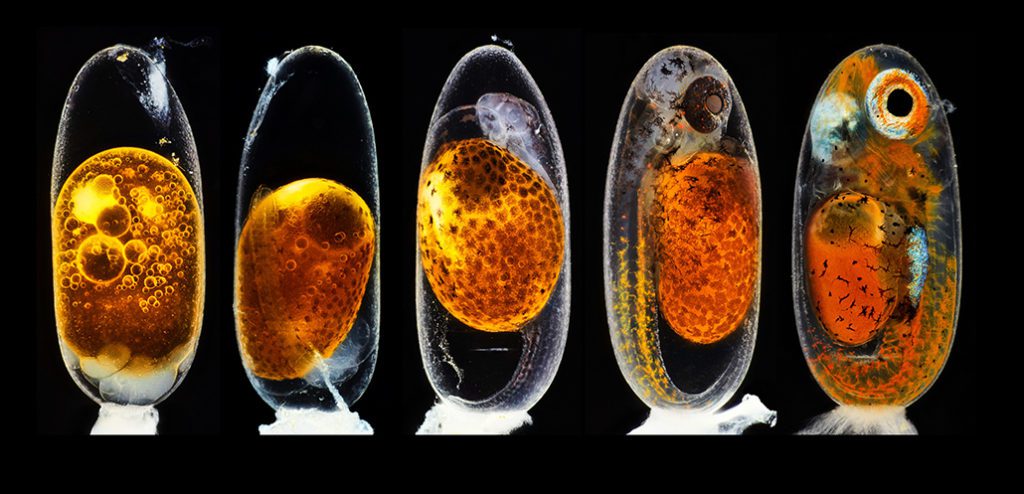
Over a period of nine days, German Photographer Daniel Knop saw an embryo transform into a baby clownfish. The five images combined into one were taken while the embryo was in motion. It shows the stages of its development from left to right. It bagged second place in the photography competition.
The first image shows the egg hours after fertilization. The next images were taken on the third day, one during the morning and second, evening. The last two images were taken on the fifth day and ninth day, hours before hatching.
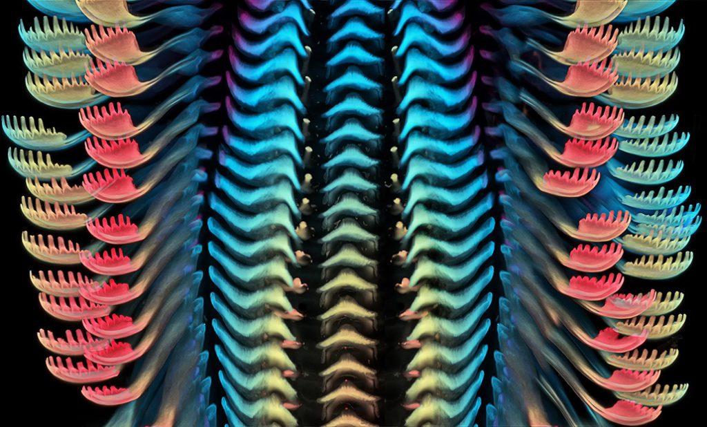
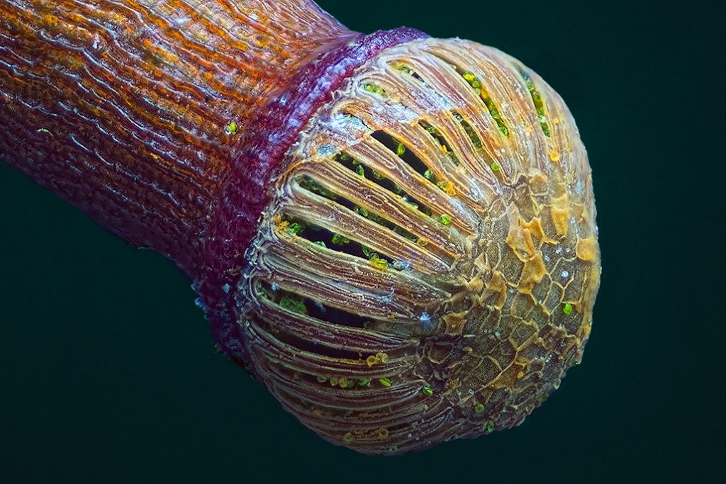
Neurobiologist Igor Siwanowicz snapped a picture of a part of a snail’s tongue when his lab mate’s aquarium was taken over by freshwater snails. This picture earned third place in the competition.
Igor Siwanowicz says, “I chose this image to show that in nature, beauty can be found in the most unexpected places, like a snail’s mouth.” He won second place in the 2019 photography competition.
Other beautiful entries
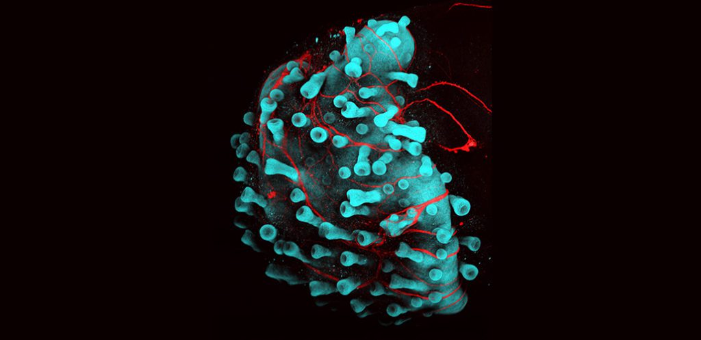
This picture was taken by Neuroscientist Karl Koehler and biochemist Jiyoon Lee. It shows human hair follicles growing off of a lab-grown cluster of skin cells, called an organoid. The blue colour shows the skin organoid, the stubby projections are hair follicles and the nerves are shown in red. The image earned an honourable mention in the competition.
This image of distinction in the photography competition was taken by Miroslav Žít, a photographer from Prachatice in the Czech Republic. It shows a moss capsule packed with spores. Once released, these spores travel a long distance through the wind.
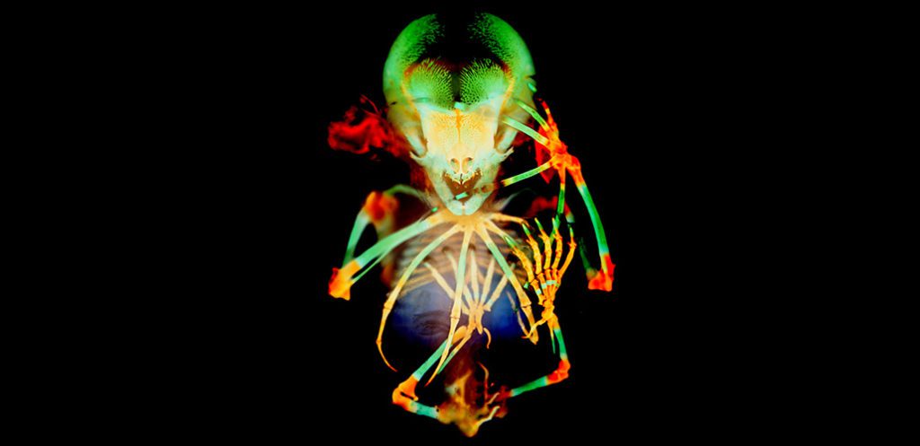
Vanessa Chong-Morrison, a developmental biologist then at the University of Oxford, prepared this image of a Seba’s short-tailed fruit bat embryo for picture day while participating in an embryology course at the Marine Biological Laboratory in Woods Hole. The bones of the bat are shown in green, and cartilage in orange. This picture secured the 20th position.
Further Reading: